
Heart tests: Types, uses, and when to contact a doctor
Th3
“We’re going to run a few heart tests.” Those words can make even the calmest person suddenly remember every questionable decision they’ve ever madelike that third espresso, the “temporary” couch lifestyle, or the time you thought stairs were optional.
Here’s the good news: most heart tests are designed to answer simple questions with as little drama as possible. Is your heart rhythm normal? Are your valves opening and closing like they’re supposed to? Is your heart getting enough blood when you’re active? Doctors use these tests to confirm what’s going on, rule out dangerous problems, and pick the right next stepwhether that’s reassurance, lifestyle changes, medication, or more specialized care.
This guide breaks down the most common heart tests, what they’re for, what they feel like, how results are interpreted, and exactly when symptoms mean you should call your doctoror call 911. (Spoiler: your heart is allowed to be quirky, but not mysteriously quirky.)
Why heart tests matter (and what they can’t do)
Think of heart testing like troubleshooting a carexcept the “car” is you, the dashboard warnings are symptoms, and the mechanic is trying to avoid unnecessary repairs while still keeping you safe. A single test rarely tells the whole story. Instead, clinicians combine:
- Your symptoms (chest pain, shortness of breath, palpitations, dizziness, fainting)
- Your risk factors (blood pressure, cholesterol, diabetes, smoking, family history)
- Physical exam and vitals
- Targeted tests that match the most likely causes
Also important: every test has limits. Some tests are great at ruling out emergencies. Others are better for long-term risk. Some can produce false alarms (a “positive” that turns out not to be a real problem), especially if your risk is low to begin with. That’s not a failureit’s just statistics being its messy self.
The big categories of heart tests
Most cardiac testing falls into a few buckets. If you know the bucket, the test suddenly sounds less mysterious and more like “oh, that one.”
1) Electrical tests: rhythm, rate, and “why is my heart doing jazz improv?”
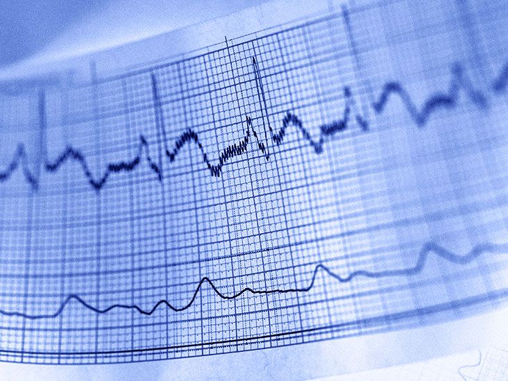
Electrocardiogram (ECG/EKG)
An ECG (or EKG) records the electrical signals that coordinate each heartbeat. Sticky electrodes go on your chest (and sometimes limbs), and the test usually takes just a few minutes.
What it’s used for: spotting abnormal rhythms (arrhythmias), signs of a past heart attack, clues to heart muscle strain, and sometimes reasons for chest pain. It’s also commonly used as a baseline before procedures or new medications.
What it feels like: like wearing stickers while a printer draws squiggles. No shocks, no pain. Your heart does all the work; the machine is just eavesdropping.
Holter monitor (24–48 hours) and event monitors (longer-term)
If symptoms are intermittentlike palpitations that show up only when you’re trying to relaxyour clinician may recommend ambulatory ECG monitoring.
- Holter monitor: continuous recording, typically 24–48 hours. Great when symptoms happen daily or near-daily.
- Event monitor: used longer (weeks), often triggered by you when symptoms occur, or automatically when the device detects an abnormal rhythm.
Why they’re helpful: they connect “I felt weird at 3:17 p.m.” with “here’s what your heart was doing at 3:17 p.m.” That timing match can be gold.
Implantable loop recorder (months to years)
For rare but concerning symptomslike unexplained faintingan implantable loop recorder can monitor rhythm for up to years. It sits under the skin on the chest and continuously checks your heartbeat, capturing events you’d never catch in a short office visit.
2) Ultrasound tests: structure, valves, and pumping power
Echocardiogram (Echo)
An echocardiogram uses ultrasound to create moving pictures of your heart. It can show chamber size, pumping function, valve movement, and fluid around the heart.
Common types:
- Transthoracic echo (TTE): the standard, noninvasive echo done on the chest wall.
- Transesophageal echo (TEE): an ultrasound probe is guided into the esophagus for clearer images (especially valves and clots). This is more invasive and usually involves sedation.
What it’s used for: valve disease, heart failure evaluation, cardiomyopathy, congenital issues, suspected blood clots, infections on valves, and more.
3) Stress tests: how your heart performs under pressure (the healthy kind)
A cardiac stress test checks how your heart behaves when it has to work harderbecause some problems don’t show up when you’re sitting calmly thinking about your inbox.
Exercise stress test (treadmill or bike)
You walk on a treadmill (or pedal) while your ECG and blood pressure are monitored. The intensity increases gradually.
What it’s used for: detecting reduced blood flow to the heart muscle (suggestive of coronary artery disease), assessing exercise tolerance, and guiding safe activity levels.
Nuclear stress test
A nuclear stress test uses a small amount of radiotracer and imaging to see blood flow to the heart at rest and during stress. This can highlight areas with reduced perfusion or prior damage.
Why it’s chosen: often when the standard exercise ECG test might be less informative, or when imaging detail is needed.
Stress echocardiogram and pharmacologic stress testing
Instead of (or in addition to) ECG changes, a stress echo compares heart ultrasound images before and after stress. If you can’t exercise adequately, medications can simulate “stress” by increasing heart workload or changing blood flow.
4) Imaging tests: arteries, plaque, and the “map of the plumbing”
Coronary calcium scan (Coronary artery calcium score)
A coronary calcium scan is a specialized CT that measures calcified plaque in coronary arteries. It’s commonly used to refine long-term risk assessment and guide prevention decisions (like whether a statin makes sense).
Key point: it’s about risk and preventionnot diagnosing an active heart attack.
CT coronary angiogram (CCTA)
A CT coronary angiogram uses CT imaging and contrast dye to look for narrowed or blocked coronary arteries. It can be especially useful for evaluating certain chest pain scenarios and providing detailed anatomy without a catheter-based procedure.
What to expect: an IV for contrast, instructions to hold your breath at times, and sometimes medication to slow the heart rate for clearer images.
Cardiac MRI
A cardiac MRI provides high-detail images of heart structure and function without radiation. It’s particularly useful for evaluating heart muscle disease, inflammation, scarring, congenital conditions, and complex anatomy.
Reality check: the scanner can be loud and enclosed. If claustrophobia is your nemesis, tell your teamthere are ways to help.
5) Blood tests: what your bloodstream can reveal about heart risk or injury
Blood tests don’t “see” the heart, but they can signal damage, strain, or future risk.
Troponin
Troponin is a protein released into the blood when heart muscle is damaged. It’s one of the cornerstone tests in evaluating suspected heart attacks, especially alongside symptoms and ECG findings.
BNP / NT-proBNP
BNP (or NT-proBNP) levels can rise when the heart is under strain, often used to help evaluate symptoms like shortness of breath when heart failure is a concern.
Lipid profile (cholesterol test)
A lipid profile measures cholesterol and triglycerides. It’s a major tool for estimating cardiovascular risk and guiding prevention.
High-sensitivity C-reactive protein (hs-CRP)
hs-CRP can reflect inflammation and may help refine risk in certain situations. It’s not a standalone “you will/won’t have a heart event” crystal ballbut it can add context.
6) Invasive tests: when doctors need direct measurement or treatment
Cardiac catheterization and coronary angiography
A coronary angiogram (via cardiac catheterization) places a thin catheter into blood vessels to inject contrast dye and visualize coronary arteries under X-ray. It can also be used to treat blockages (angioplasty/stenting) during the same procedure when appropriate.
When it’s used: higher-risk chest pain, suspected significant coronary disease, abnormal noninvasive test results, or situations where intervention may be needed.
Electrophysiology (EP) study
An EP study evaluates the heart’s electrical pathways using catheters and detailed mapping. It’s used to diagnose and sometimes treat certain arrhythmias (for example, by performing an ablation during the same session).
7) Specialty tests for specific symptom puzzles
Tilt table test
If you faint or nearly faint and the cause isn’t obvious, a tilt table test can help. You’re strapped safely to a table that tilts from lying to upright while vitals are monitored. The goal is to see how heart rate and blood pressure respond to posture changes.
Cardiopulmonary exercise testing (CPET)
CPET measures how the heart and lungs perform during exercise by analyzing breathing, oxygen use, and heart rhythm. It’s especially helpful when shortness of breath has multiple possible causes and you need a clearer “who’s responsible here?” answer.
How doctors decide which test you need
Choosing a heart test isn’t about ordering the fanciest machine. It’s about matching the test to the question.
- Chest pain with concern for a heart attack: ECG + troponin are usually front-line.
- Palpitations: ECG first, then Holter/event monitor if symptoms come and go.
- Shortness of breath or swelling: BNP/NT-proBNP and an echocardiogram often enter the chat.
- Risk assessment without symptoms: lipid profile is foundational; coronary calcium scan may be considered for risk refinement in selected people.
- Possible coronary blockages: stress testing, CCTA, or catheterization depending on risk and context.
- Fainting: rhythm monitoring, tilt table testing, and sometimes EP studies if indicated.
If you ever feel like the plan is “test roulette,” it’s fair to ask: “What question are we trying to answer, and how will the result change what we do next?” Good clinicians love that question. It keeps testing purposeful.
What to expect and how to prepare (without going full Google-doom)
Preparation varies by test, but a few themes repeat:
- Medication instructions: Some tests require holding certain meds (like beta blockers) while others do not. Follow your clinician’s guidance rather than self-adjusting.
- Caffeine rules: Some stress tests and tilt tests ask you to avoid caffeine beforehand.
- Fasting: Imaging with contrast or certain procedures may require fasting for a set time.
- Comfort: Wear easy clothing and shoes you can exercise in if it’s a treadmill test.
- Allergies and kidney history: Important for tests using contrast dye.
- Implants/metal devices: Essential to mention before MRI.
And yes, you can ask how long it takes. Not because you’re impatient, but because you have a life and your heart isn’t the only thing with a schedule.
Understanding results: normal, abnormal, and “could be nothing, could be something”
Results are rarely just “good” or “bad.” Many are “this finding might matter in the right clinical context.” Here’s how to keep your sanity:
Abnormal doesn’t always mean dangerous
For example, a harmless premature beat can feel dramatic but be medically low-risk. Mild valve leakage is common. Some ECG “abnormalities” are normal variants for certain people.
Normal doesn’t always mean “nothing is wrong”
A normal ECG can happen between episodes of arrhythmia. A normal stress test reduces the likelihood of major blockages but doesn’t erase all risk forever. That’s why clinicians pair tests with symptoms and risk profiles.
False positives happen
No test is perfect. In lower-risk people, some tests can flag something that turns out not to be clinically important. This is another reason why the “what question are we answering?” approach matters.
When to contact a doctor (and when to call 911)
This section matters most. If you remember nothing else, remember this: it’s better to be safely evaluated than to tough it out in silence.
Call 911 (or your local emergency number) right away if you have:
- New, unexplained, severe, or persistent chest pain (especially pressure, squeezing, fullness, or pain lasting more than a few minutes, or coming and going)
- Chest discomfort plus shortness of breath, sweating, nausea, lightheadedness, or fainting
- Pain spreading to the arm(s), back, neck, jaw, or upper stomach
- Severe shortness of breath at rest or worsening rapidly
- Fainting (syncope), especially with chest pain or palpitations
- Sudden weakness, trouble speaking, or facial droop (stroke symptomsstill a 911 situation)
Contact your doctor soon (same day or within a few days) if you have:
- New or worsening palpitations, especially if they’re frequent or last longer than usual
- Dizziness or near-fainting
- Shortness of breath with activity that’s new or clearly worsening
- Swelling in legs/feet, sudden weight gain, or fatigue that feels out of proportion
- Chest discomfort with exertion that improves with rest (even if it’s mild)
Important: If you have symptoms that feel urgent, don’t try to “earn certainty” by waiting for a test appointment. Emergencies don’t RSVP.
Conclusion
Heart tests are toolssmart tools, often surprisingly simple toolsthat help clinicians translate symptoms and risk into clear decisions. Some tests focus on rhythm (ECG, monitors), some on structure (echocardiogram), some on performance (stress tests), some on arteries and plaque (CT-based tests), and some on biochemical clues (troponin, BNP, lipid profiles). The best testing strategy isn’t “do everything,” but “do what answers the right question.”
If you’re ever unsure, use this rule of thumb: new chest pain, severe shortness of breath, fainting, or symptoms that feel “wrong” in a serious way = emergency evaluation. For ongoing, recurrent, or worsening symptoms, your doctor can guide you to the most appropriate heart testwithout turning your calendar into a medical mini-series.
Real-world experiences: what people commonly notice before, during, and after heart tests (about )
Let’s talk about the part nobody puts on the brochure: the human experience of heart testing. Not the lab valuesyour thoughts, your nerves, and the oddly specific things you’ll remember later.
The “I thought I was fine… until I had to lie still” moment
Many people feel okay walking into the appointment, then suddenly become hyper-aware of every heartbeat once the electrodes go on. It’s normal. When you’re told “don’t talk, don’t move,” your brain hears: “Now is the perfect time to obsess.” Pro tip: slow breathing helps, and so does remembering the ECG is measuring younot judging you.
Stress tests: equal parts science and “why is this treadmill so honest?”
People often describe exercise stress tests as less scary and more awkwardly motivational. The pace ramps up, and you learn two things quickly: (1) your legs have opinions, and (2) the staff has seen every version of “I’m fine” ever performed by a sweating human. Some folks worry they’ll “fail” the test. You can’t fail. The goal is to gather information safely, not to crown the treadmill champion of Tuesday.
Echocardiograms: surprisingly chill, surprisingly gooey
Transthoracic echoes usually feel like a calm spa dayexcept the spa uses gel that will somehow end up on your shirt no matter what you do. People are often fascinated seeing their heart move in real time. It’s one of the few moments in modern life where you can literally watch yourself function.
Monitors: the sticker saga
Holter and event monitors tend to create two types of experiences: “This is no big deal” and “Why do these stickers have the grip strength of a thousand suns?” Some people get mild skin irritation from adhesive. If you’re sensitive, ask about skin prep tips or alternative options. Also: keeping a symptom diary feels old-school, but it can be extremely helpfulespecially if your palpitations only show up during three specific situations: work meetings, family group chats, and the moment you finally fall asleep.
CT and MRI: the waiting is the hardest part
Imaging tests often come with anticipatory anxiety. Many people say the scan itself wasn’t badthe waiting for results was the worst part. If you’re stuck in “what if” spirals, it can help to ask upfront when you’ll hear back and what the next steps might be in each scenario. A plan doesn’t eliminate worry, but it gives worry fewer places to hide.
After invasive procedures: relief plus a weird new respect for your body
People who undergo catheterization or EP studies often describe a mix of relief (“we finally know what it is”) and awe (“modern medicine is wild”). It’s also common to feel tired afterward, especially if sedation was used. The most consistent advice from patient experience: follow the post-procedure instructions exactly, and don’t try to prove you’re tough by doing too much too soon. Your heart is already doing the heavy lifting. Let it win the argument.